
Initial Management of Chemical Burns
Henrietta Creasy and Peter Drew
0.50 Hours
This session will describe how to decontaminate the skin following chemical exposure, focusing in depth on the initial treatment of hydrofluoric acid and phosphorus burns.
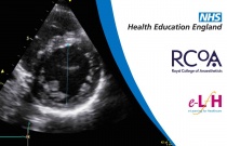
Left Ventricular Function
Dave Woodward
0.50 Hours
This session will explain normal left ventricular (LV) function and how to diagnose an abnormal left ventricle (LV). Qualitative and quantitative methods of assessing LV function will be described. Attention is drawn to ways in which pathology and interventions may influence the interpretation of a basic transthoracic echocardio....
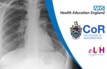
Image Interpretation - Plain X-rays of the Adult Chest: Interstitial Lung Disease
Andrew Yeung
This session will look at the interstitial pattern of opacification which is commonly seen in chest radiographs. The anatomy and physiology of the lung interstitium will be presented. This will be correlated with the clinical and radiological features associated with common interstitial lung diseases, and other processes, which....

Image Interpretation - Radiographs of the Adult Abdomen: Normal Abdominal Radiograph
Nigel Hughes
0.50 Hours
This session will look at the normal radiograph of the abdomen. It will focus on identifying anatomy and the normal bowel gas pattern demonstrated on radiographs. Indications for abdominal radiography will be discussed along with guidance on writing a report.

Image Interpretation - Radiographs of the Adult Abdomen: Gas and Soft Tissue Abnormalities
Andrew Yeung
0.50 Hours
This session will look at the gas and soft tissue structures of the abdomen and how they appear on abdominal radiographs. Common pathological processes in relation to these are presented and key imaging findings are described.

Gastrointestinal Complications of Burns
Anne-Marie Kennedy and Odhran Shelley
0.50 Hours
This session will describe the gastrointestinal complications of burns and explain the aetiology of peptic ulceration and ileus in patients with burns. Later, it will describe how to avoid, recognise and treat such gastrointestinal complications.

Importance of Homeostatic Control
Alan Noble
0.50 Hours
Maintenance of homeostasis, the constancy of the internal environment, is essential for the continuation of life. In this session some fundamental concepts are introduced and the key closely controlled physiological parameters are identified.

General Surgery Abdominal Swelling
Polly Estridge
0.50 Hours
This session aims to give an overview of the aetiology of abdominal masses and consider how they might present. It will provide a structured approach to diagnosis and a general strategy towards management of these patients.

General Surgery Change in Bowel Habit
Hannah Knight
0.50 Hours
This session will cover the surgical principles associated with a change in bowel habit including definitions, aetiology, assessment and investigation.

Genitourinary Disease: Haematuria
Madeline Moore
0.50 Hours
This session will provide an overview of haematuria including when it’s significant, its causes and initial investigations and management.

Endocrine: Acute Endocrine Crises
Constantine Halkias and Vas Constantinides
0.25 Hours
This session looks at the importance of the surgical trainee to be able to recognize, formulate a treatment plan and select surgical intervention for acute endocrine crises.

General Paediatric Trauma - Special Considerations
Kevin Cao and Alexander Alexiou
0.50 Hours
An introduction to important considerations in Paediatric trauma as well as considerations in assessing patients with non-accidental injuries.

Fractures: Classification of Fractures
Efthymios Iliopoulos
0.50 Hours
This session will review the most commonly used classifications of fractures and describe their clinical relevance.

Management of Blood Glucose
Jan Idkowiak
0.50 Hours
This session outlines the importance of regular insulin therapy, blood glucose monitoring, and the role of diet in the management of type 1 diabetes.

IUT: Managing Unexpected Situations and Complications
John Bland
0.50 Hours
This session identifies problems which may arise during insertion of intrauterine contraceptive methods and describes how to manage those situations.

Local Anaesthesia for Subdermal Implant Insertion and Removal
Liz Stephens
0.50 Hours
This session explores the principles of local anaesthetic use for subdermal implant (SDI) insertion and removal.

Genital Ulcerations
Tatiana Tchikhiaeva and Martin Talbot
0.50 Hours
This session provides an overview of the common causes of genital ulceration, their diagnosis and basic management.

Mechanism of Action and Contraceptive Effectiveness
Sam Rowlands
0.50 Hours
This session covers modes of action of contraceptives, distinctions between reversible and permanent methods, differences in discontinuation rates between methods and how contraceptive failure is measured.

Drug Addiction, Dependency and Pain - Management
James Bell
This session describes the key concepts of the management of pain in patients with a history of substance misuse

Heuristics and Bias
Kerry Crawley
0.50 Hours
Heuristics are defined as ‘mental shortcuts’, abbreviated decision making’ processes, or rules that allow us to simplify complex decisions.

Hypothetico-deductive Decision Making
Kerry Crawley
0.50 Hours
Along with pattern recognition, the hypothetico-deductive method is the most prevalent mode of decision making in emergency care (Xu et al.2012).

Drugs Acting on the Respiratory System
John Kinnear
0.50 Hours
This session looks at drugs which are used primarily for their effects on the respiratory system, with particular emphasis on those drugs that may be encountered by the anaesthetist. Therapies for the management of acute asthma are only briefly mentioned since they will be explored in greater detail in Module 07c/Systematic Phar....

Drugs Used in the Treatment of Acute Asthma
John Kinnear
0.50 Hours
This session considers the management of acute asthma from a mainly pharmacological perspective. A clinical case is described for illustrative purposes.

Newtonian Mechanics
Elke Kothmann and Kaye Cantlay
0.50 Hours
The aim of this session is to consider the concepts of force and mass and to introduce Newton’s three laws of motion.

Gases in Solution
Rahul Bajekal
0.50 Hours
This session looks at some physical principles behind gas solubility and their clinical applications.